
Radiography & Radiology Explained By A Local Glendale Doctor
The field of radiology uses medical imaging to identify and cure disorders that affect the human body. When German physicist Wilhelm Conrad Röntgen discovered X-rays in 1902, radiology was first used. Since then, radiography has advanced significantly, moving from early films that took hours to prepare to contemporary CT scans that produce in-depth images in minutes. To diagnose or treat a wide range of disorders, imaging techniques including X-ray radiography, ultrasound, computed tomography (CT), nuclear medicine, including positron emission tomography (PET), and magnetic resonance imaging (MRI) are utilized. As new diagnostic and therapeutic techniques are created, the field of radiology continues to develop. In more recent years, radiology has expanded to include diagnosis using an organ-scanning technique that combines the use of radioactive isotopes and nonionizing radiation, such as ultrasound and nuclear magnetic resonance. Similar to how hormones and chemotherapeutic medications are now included in the spectrum of radiotherapy for the treatment of cancer. In this article, a local Glendale, Queens Radiologist will build us a complete picture of everything you need to know about Radiology.
WHAT IS RADIOLOGY?
Radiology is a branch of medicine that uses medical imaging to diagnose and treat various conditions and diseases. It plays an important role in the medical industry, as it helps doctors to get a better understanding of what is going on inside the human body. The modern practice of radiology involves several different healthcare professionals working as a team.
A physician with the necessary post-graduate training in radiology, a radiologist evaluates medical images, reports his or her findings to other doctors verbally or in writing, and employs imaging to carry out minimally invasive medical operations. The nurse is involved in the treatment of patients both before and after scans or treatments, and this includes administering drugs, keeping an eye on patients who are sedated, and monitoring vital signs. There are numerous forms of radiology, and each one offers a special set of advantages.
WHAT ARE THE DIFFERENT TYPES OF RADIOLOGY?
Radiology is divided into two different areas, diagnostic radiology and interventional radiology. Doctors who specialize in radiology are called radiologists.
- DIAGNOSTIC RADIOLOGY: Diagnostic radiography is a subspecialty of medicine that makes use of imaging methods including X-rays, CT scans, and MRIs to make medical diagnoses. These tests’ images can be used to help doctors identify the location and severity of a patient’s injury or sickness and suggest appropriate courses of action. One of the most crucial instruments in contemporary medicine is diagnostic radiology. It aids medical professionals in locating disorders with internal organs including the kidneys, brain, heart, and lungs as well as in spotting anomalies in bones and other tissues.
Diagnostic imaging also gives medical professionals the ability to direct minimally invasive operations that include the insertion of surgical equipment or needles into the body through tiny incisions. Patients’ entire quality of life can be enhanced while also experiencing less pain and a faster rate of recovery. The radiologist or other medical professionals can frequently:
- diagnose the source of your symptoms using the diagnostic images,
- monitor how well your body is responding to a treatment you are receiving for your disease or condition,
- and screen for various illnesses like breast cancer, colon cancer, or heart disease.
The most common types of diagnostic radiology exams include:
- Computerized axial tomography (CAT) scans, usually referred to as computed tomography (CT), which includes CT angiography
- Fluoroscopy, including barium enema and upper GI
- Magnetic resonance angiography (MR angiography) and magnetic resonance imaging (MRI) (MRA)
- Mammography
- Nuclear medicine, which uses procedures including a thallium cardiac stress test, thyroid scans, and bone scans.
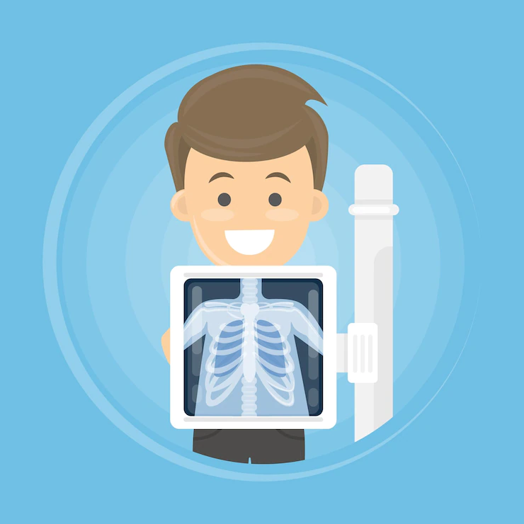
- Plain radiographs, such as chest radiographs
- Positron emission tomography, sometimes referred to as PET imaging, PET scanning, or PET-CT when used in conjunction with CT
- Ultrasound
- INTERVENTIONAL RADIOLOGY: Medical professionals known as interventional radiologists use imaging techniques like CT, ultrasound, MRI, and fluoroscopy to help direct treatments. When placing catheters, wires, and other tiny equipment and instruments into your body, the doctor can use the imaging to aid. Smaller incisions may usually be made due to this (cuts).
Instead of seeing directly inside your body with a scope (camera) or with open surgery, doctors can utilize this technology to diagnose or treat diseases in nearly any part of the body. In addition to treating back pain, liver issues, renal issues, uterine fibroids, artery and vein obstructions, and malignancies or tumors, interventional radiologists also frequently treat these conditions. No incision, or just a very small one, will be made by the doctor. Rarely is it necessary for you to remain in the hospital after the procedure. In most cases, just mild sedation is necessary (medicines to help you relax).
Interventional radiology techniques include, for instance:
- Angiography or angioplasty with stent placement
- Embolization to stop bleeding
- Cancer treatments such as tumor embolization using chemoembolization or Y-90 radioembolization
- Tumor ablation with radiofrequency ablation, cryoablation, or microwave ablation
- Vertebroplasty and kyphoplasty
- Needle biopsies of various organs, such as the lungs and thyroid gland
- Breast biopsy, guided either by stereotactic or ultrasound techniques
- Uterine artery embolization
- Feeding tube placement
- Venous access catheter placement, such as ports and PICCs
WHAT ARE THE BENEFITS OF RADIOLOGY?
Radiology is a powerful tool that can be used to diagnose and treat a wide range of medical conditions. Here are 10 reasons why radiology is so important:
- Radiology allows doctors to see inside the body without having to perform surgery, which can be dangerous and expensive.
- Radiology helps doctors detect early signs of disease, so they can start treatment sooner and prevent the development of more serious conditions.
- Radiology reduces the need for invasive procedures that may cause long-term damage or complications, such as catheterization or biopsies.
- Radiology helps doctors determine if there are any complications during procedures like surgeries or MRIs/CT scans, so they can adjust their treatment plan accordingly.
- Radiology enables doctors to better understand existing diseases so they can make informed decisions about treatment options and work with patients on an individual basis to develop a plan that meets each person’s unique needs (e.g., diet changes).
- Radiology helps doctors identify risk factors that could lead to future problems like heart disease or cancer so they can provide guidance on how these issues should be addressed moving forward (e .g., exercise more, eat healthier, stop smoking).
- Radiology helps doctors spot potentially dangerous conditions like aneurysms and tumors so they can quickly alert patients before they cause any serious damage to the body.
- Radiology allows doctors to work with their patients as they develop treatment plans that will address any issues that have been identified through diagnostic imaging so they can prevent complications from occurring in the future (e.g., radiation oncology).
- Radiology is also used to monitor the effectiveness of treatments that have been prescribed by doctors so they can determine if adjustments need to be made for better results.
- Finally, radiology helps doctors diagnose problems in the form of cancerous tumors (e .g., breast or prostate) and other conditions that may require early intervention and treatment before they become life-threatening.
RADIOGRAPHY
An imaging method called radiography uses X-rays, gamma rays, or other ionizing radiation along with non-ionizing radiation to see an object’s internal structure. Medical radiography (“diagnostic” and “therapeutic”) and industrial radiography are two examples of applications for radiography. Airport security employs similar methods (where “body scanners” generally use backscatter X-ray). In traditional radiography, a beam of X-rays is projected toward the object in order to make an image. Depending on the object’s density and structural makeup, a specific amount of X-rays or other radiation is absorbed by it. A detector is placed behind the object to collect the X-rays that travel through it (either photographic film or a digital detector).
DIFFERENCES BETWEEN RADIOGRAPHY AND RADIOLOGY
- Radiography is a medical technology, whereas radiology is a specialist area of medicine.
- Radiography is the process of creating images of body organs that serve as the foundation for radiologists in the diagnosis and treatment of certain ailments. Radiology is the diagnosis and treatment of diseases.
- Radiology requires more time to study because only individuals with a medical degree can pursue a career in it, whereas radiography only requires a high school education.
- Radiography makes use of technologies including MRI, ultrasound, mammography, CT scan, x-ray, and nuclear medicine to capture crisp images that are subsequently examined by radiologists.
APPLICATIONS OF RADIOLOGY IN HEALTH CARE
Radiology is a field of medicine that deals with the use of X-rays to diagnose and treat health issues. It’s used in many areas of healthcare, including:
- Radiation oncology: Radiation therapy kills cancer cells in cancerous tumors to treat them (or shrinking them). By bringing together the most recent findings and advancements in the field, Radiation Oncology offers an open access forum for academics and medical professionals working to manage and treat cancer patients.
- Nuclear medicine: To help diagnose specific illnesses, nuclear medicine injects minute amounts of radioactive material into the body (such as heart disease). The natural environment exposes us to ionizing radiation every day, but additional exposures, such as those from nuclear medicine treatments, can modestly raise the chance of acquiring cancer in later life.
- Bone densitometry: This examination gauges bone density and can be used to identify osteoporosis. Millions of women are at risk for potentially disabling fractures due to osteoporosis, which causes a considerable reduction in bone mineral density. In order to determine a patient’s fracture risk, we provide bone densitometry, which precisely analyzes bone mineral density (BMD). Our sophisticated method can determine BMD in the wrist, hip, or spine with accuracy. The technique also allows for the measurement of BMD in children.
- Radiation therapy: Radiation is used to treat malignant tumors by reducing their size. As a result of its capacity to regulate cell proliferation, radiation treatment is frequently administered to malignant tumors. Ionizing radiation kills cells by destroying the DNA of malignant tissue. Shaped radiation beams are directed from different exposure angles to intersect at the tumor, giving it a considerably higher absorbed dose than the surrounding healthy tissue in order to spare normal tissues.
- Magnetic resonance imaging (MRI): This is a noninvasive test that creates images of organs and other structures within your body using powerful magnets, radio waves, and computer technology. is a medical imaging technique used in radiology to form pictures of the anatomy and the physiological processes of the body. MRI scanners use strong magnetic fields, magnetic field gradients, and radio waves to generate images of the organs in the body. MRI does not involve X-rays or the use of ionizing radiation,
- Computed tomography (CT scan): refers to a computerized x-ray imaging procedure in which a narrow beam of x-rays is aimed at a patient and quickly rotated around the body, producing signals that are processed by the machine’s computer to generate cross-sectional images, or “slices.” These slices are called tomographic images and can give a clinician more detailed information than conventional x-rays.
ARE THERE ANY RISKS ASSOCIATED WITH RADIOLOGY?
Radiology is a highly specialized field of medicine that requires extensive training and knowledge to practice safely. The risks associated with radiology are significant, and they must be understood by all medical professionals who work in this field.
The following are some of the most common risks associated with radiology:
- Exposure to ionizing radiation (X-rays)
- Exposure to radioactive materials
- Accidents or mistakes that occur during an examination, such as an incorrect injection or wrong patient identification
- Injuries from equipment used during the examination (such as broken bones)
- Injuries from working in a cramped space and moving heavy equipment, such as back injuries
- Burns or cuts if any part of the body comes into contact with hot equipment or radioactive material during an exam
- Errors in interpreting the images created by radiological procedures, which could lead to misdiagnosis or improper treatment
CONCLUSION
Radiology is an important field in the medical industry. It helps doctors to diagnose and treat patients. There are many different types of radiology, and each has its own unique benefits. Radiologists play a vital role in the medical industry, and they will continue to do so for many years to come. The primary goal of radiology is to create an image that allows a radiologist to discern as much information about a person’s health as possible. This includes identifying any abnormalities or injuries in the body, determining their severity, and recommending treatment plans for patients.
Contributed by: EMU Radiology Center Queens 8340 Woodhaven Blvd Ste 7 Glendale, NY 11385 929-299-6126