
Glendale Workers Face Rising Injury Rates in Queens
By Dan Rose,
Something has shifted in Glendale over the past few years, and the numbers tell the story before anyone else does. I have watched the volume of workers’ compensation cases coming out of this neighborhood climb steadily, and the pattern is not random. It tracks directly with the changes happening on Glendale’s streets, in its warehouses, and across its evolving workforce.
Glendale has always been a working neighborhood. Its identity was built by factory hands, tradespeople, and small-business owners who kept their heads down and got the job done. But the scale of activity happening here now is different from what the neighborhood handled a generation ago. ZIP code 11385 is one of the most densely populated in New York State, with a population exceeding 100,000 residents crammed into a few square miles. That density ripples into every workplace, every commercial corridor, and every construction site in the area.
Why Glendale’s Workplaces Are Getting More Dangerous
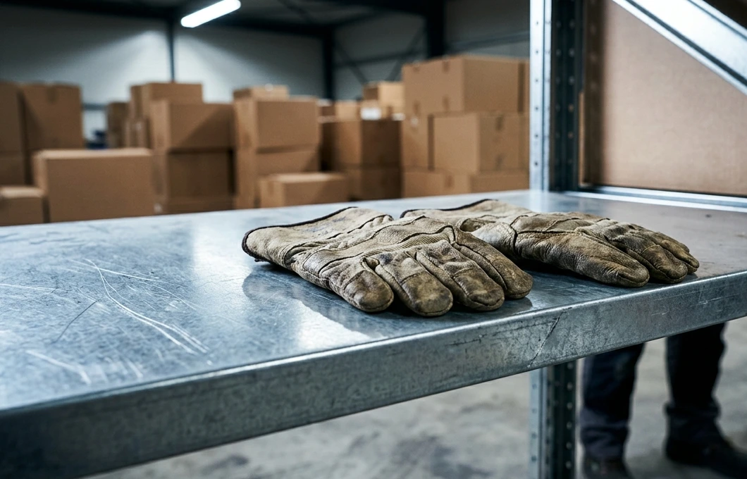
The surge in claims is not coming from one industry. It is spread across the sectors that define Glendale’s economy. Warehouse and distribution operations along Cooper Avenue and near the old Atlas Terminal corridor have expanded aggressively to keep pace with e-commerce fulfillment demands. These facilities run fast, sometimes around the clock, and the pressure to move product quickly leads to shortcuts. Forklifts operating in tight aisles. Workers lifting loads without proper equipment. Inadequate training for new hires who barely have time to learn the layout before they are expected to keep pace with veterans.
Construction is another major driver. Glendale’s housing stock dates overwhelmingly to the pre-war era, with more than 70 percent of residential buildings constructed before 1940. Renovation and new infill development are happening on virtually every block, and these projects bring the full range of construction hazards. Falls from scaffolding, struck-by incidents, caught-between injuries involving heavy equipment on cramped lots where there is barely room to maneuver.
Then there are the jobs that don’t make headlines but generate a surprising volume of claims. Restaurant workers along Myrtle Avenue suffering burns, repetitive strain injuries, and slip-and-fall incidents in kitchens built decades before modern safety codes existed. Retail employees at shopping centers lifting inventory and standing on concrete floors for ten-hour shifts. Home health aides traveling between clients and performing physically demanding patient care with little institutional support.
Congestion, Growth, and Diminished Safety Standards
Part of what makes Glendale’s situation distinct is the collision between rapid growth and limited physical space. This is not a neighborhood with wide boulevards and sprawling industrial parks. Workplaces here are stacked into older buildings, squeezed onto narrow lots, and sandwiched between residential streets where delivery trucks compete with pedestrians for every inch of pavement.
That congestion creates cascading safety problems. Loading docks designed for a fraction of today’s traffic volume now handle constant deliveries. Construction staging areas spill onto sidewalks. Workers move between tasks in spaces that were never engineered for the intensity of use they are getting now. And when an employer is trying to keep overhead low in one of the most expensive real estate markets in the country, safety measures are often the first thing to get trimmed.
Glendale, NY lawyers have recently reported a tremendous uptick of work injuries tied directly to these conditions. Congested workplaces, diminished safety standards, and a workforce that includes a significant number of undocumented or informally employed individuals who may not know their rights under New York’s no-fault workers’ compensation system. That last point matters enormously, because New York law covers virtually every employee regardless of immigration status. But if nobody tells a worker that, the injury goes unreported, the claim never gets filed, and the employer faces no accountability.
What Injured Glendale Workers Need to Know Right Now
If you have been hurt on the job in Glendale, the clock starts immediately. New York requires written notice to your employer within 30 days of the injury, and your formal claim with the Workers’ Compensation Board must be filed within two years. For injuries that develop gradually, like carpal tunnel syndrome or chronic back conditions from repetitive lifting, those deadlines run from the date you knew or should have known the condition was work-related.
The maximum weekly benefit for lost wages currently sits at $1,222.42 for injuries occurring through June 30, 2026. That rate is locked to your date of injury and will not increase even if future caps go higher. Getting your claim on file early and with proper medical documentation is essential, because insurance carriers in this market are aggressive about challenging claims, especially repetitive stress injuries where the connection to work duties is not immediately obvious.
Here is what I tell every Glendale workers’ compensation client in Queens who walk through our office door. Document everything from day one. Put your injury report in writing. See a physician who understands how to document occupational injuries. And talk to an attorney before the insurance company starts shaping the narrative of your case.
- Report in Writing: A verbal mention to your supervisor is not enough. Written notice creates a record the carrier cannot dispute later.
- Medical Records Matter: Your treating physician’s notes need to draw a clear line between your condition and your daily job duties.
- Know Your Worth: Benefits cover medical treatment, lost wages, and potential permanent disability awards. Do not settle before you understand the full scope of what you are owed.
Glendale’s workers built this neighborhood with their hands, their backs, and their persistence. When the job takes a physical toll, the law exists to make sure they are not left covering the cost alone.
Contributed by Dan Rose, A Senior Workers’ Compensation and Workplace Injury Analyst.
Need Legal Help After a Glendale Workplace Injury?
The team at Beck Law P.C. has recovered millions for injured New Yorkers and offers free, no-obligation case evaluations.
Visit us at https://becklawny.com/ to connect with an experienced workers’ compensation attorney today.
Get Directions Below!
Beck Law, 71-19 80th St Suite 8-206, Glendale, NY 11385, (516) 388-7785